Figure 1.

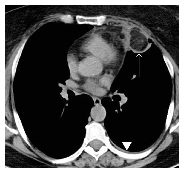
CT scan without contrast enhancement showing a left anterior low-attenuation (−80 HU) pericardial mass with high attenuation strands surrounded by an irregular capsule (arrow) and a small pleural effusion (arrowhead).

CT scan without contrast enhancement showing a left anterior low-attenuation (−80 HU) pericardial mass with high attenuation strands surrounded by an irregular capsule (arrow) and a small pleural effusion (arrowhead).