Figure 6.
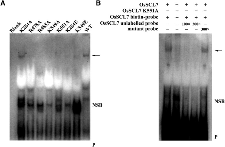
EMSA Analyses of Os-SCL7 and Model-Defined Oligonucleotide.
(A) In EMSA analyses with same amounts of probe, a band shift (arrow) can be observed for Os-SCL7 and mutant K284A, but not for other mutants. Other Os-SCL7 mutants caused a dramatic reduction in binding.
(B) EMSA competition analysis. Biotin-labeled oligonucleotide probe incubated with Os-SCL7, or mutant K551A as a negative control, in the absence or presence of 100- and 300-fold molar concentration of the unlabeled probe and competitive mutant oligonucleotide probe, respectively. Arrow, DNA binding protein; NSB, nonspecific binding; P, free biotin-labeled probe.

