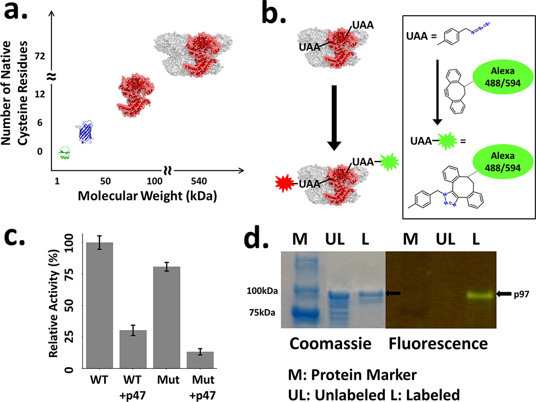
Figure 1.

(a) Size comparison of p97 and other proteins studied by site specific labeling and smFRET. CI2 (Green, PDB ID 1YPC), the first 2-state protein studied by smFRET. GFP (Blue, PDB ID 1EMA), commonly used model protein for unnatural amino acid incorporation. p97 monomer and hexamer (Red/Gray, PDB ID 3CF1), the monomer is colored red. (b) Schematic of fluorophore labeling of a multi-unnatural amino acid p97 mutant with copper-free ‘click’ chemistry. (c) ATPase activity assay of p97 wild-type (with C-terminal His-6 tag) and mutant (F131Q382) in the absence or presence of p47. Error bars indicate twice the standard deviation of the measured activities. (d) SDS-PAGE for Alexa Fluor 488 labeled p97 (F131Q382). Coommassie stain (left) and fluorescence (right, excitation wavelength 450 nm). Multiple bands in unlabeled protein lane indicate impurities, which are removed during further purification of labeled protein. Only the relevant portion of the gel is shown. For original images, see Supporting Information, Figure S1.
