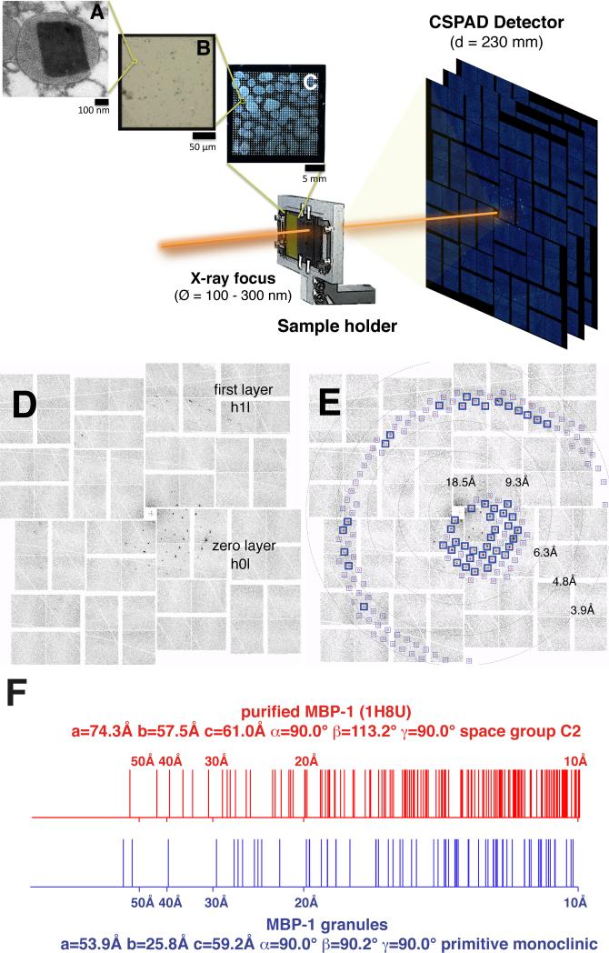
Figure 1. Crystalline Nature of MBP-1 In Its Granule Environment.
(A–C) Schematic of the experimental setup using XFEL in combination with fixed targets. The diameter of the X-ray beam (Ø) at the focal point was 100–300 nm full width at half maximum, whereas the sample-to-detector distance (D) was 230 mm. (A) TEM micrograph of a section of a resin-embedded granule showing an MBP-1 nanocrystal. (B) Magnification of a single 200 × 200 μm silicon nitride window surrounded by a silicon frame. Thousands of intact isolated granules are deposited on the surface. (C) A complete wafer.
(D) Diffraction patterns obtained when the XFEL beam intercepts a single MBP-1 nanocrystal within its cellular environment.
(E) Overlay of the observed diffraction spots with the predictions of the calculated lattice constants (blue boxes). The excellent agreement validates the lattice determination. Blue boxes highlight the predictions fulfilled by experimental data.
(F) Comparison of observed diffraction with lattice spacings of purified and recrystallized MBP-1 illustrating the differences between the two structures. Vertical lines mark the Bragg spacings (10–100 Å resolution) for two types of crystals: intragranular MBP-1 nanocrystals calculated from XFEL diffraction patterns and MBP-1 purified and recrystallized in vitro as calculated from PDB ID code 1H8U.

