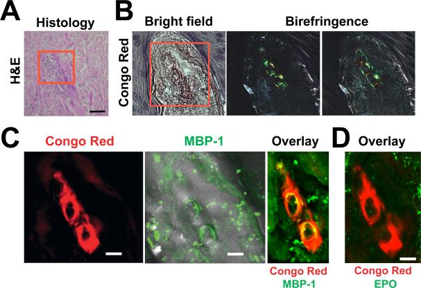
Figure 5. Extracellular MBP-1 Amyloid Deposits in Eosinophilic Tissue.
(A) Skin biopsy from a Wells’ syndrome patient. Histology shows typical collagen bundles; i.e., flame figures (red box). Scale bar, 100 μm.
(B) The area contains amyloids stained by Congo red that appear pale red in bright-field mode. When visualized between crossed polarizer and analyzer, yellow-green birefringence was present.
(C) Immunofluorescence analysis showed large extracellular MBP-1 deposition that partly overlapped with the CR fluorescence.
(D) In contrast, no EPO protein was detected.

