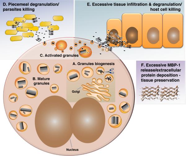
Figure 6. A Model For the MBP-1 Self-Association Cycle.
(A) MBP-1 condensation begins before/at the same time of pro-protein processing in granules. The process is followed by compaction of the cores. Granule biogenesis occurs during eosinophil differentiation.
(B) A number of mature specific granules with a crystalline interior are dispersed in the cytosol.
(C) Following eosinophil activation, the granule pH drops, and the content is mobilized, unpacked, and released through secretory vesicles.
(D) MBP-1 exerts its antibacterial effect through aggregation.
(E) MBP-1 toxicity toward host cells is also mediated by aggregation.
(F) Under conditions of sustained activation and massive secretion of MBP-1, extracellular deposition of MBP-1 can take place. Parallel arrows indicate amyloid aggregates, scissors represent the unknown protease processing pro-MBP-1, and active and toxic MBP-1 are shown as gray ellipses.

