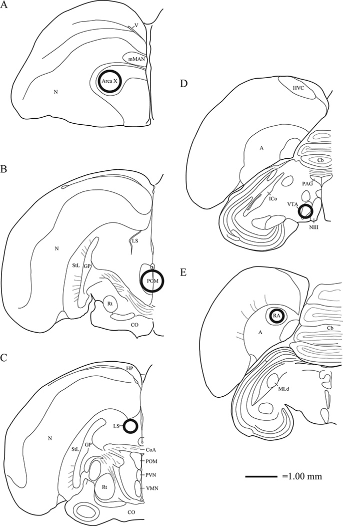
Figure 4.
Location of tissue punches illustrated in one, coronal hemisphere of the starling brain. Punches were taken bilaterally, except in POM for which a single central punch was collected. Sections progress rostrally to caudally from A – E. Approximate punch sizes and locations are represented by circles centered in Area X and POM (1.25 mm diameter), LS, VTA, and RA (0.75 mm diameter). Abbreviations listed alphabetically: A = arcopallium; Cb = cerebellum; CO = optic chiasm; CoA = anterior commissure; GP = globus pallidus; HP = hippocampus; HVC = used as a proper name; ICo = nucleus intercollicularis; MLd = mesencephalicus lateralis, dorsalis mMAN = medial portion of the magnocellular nucleus; N = nidopallium; NIII = 3rd cranial nerve; PAG = periaqueductal gray; PVN = periventricular nucleus; Rt = nucleus rotundus; StL = striatum laterale; V = ventricle; VMN = ventromedial nucleus of the hypothalamus.

