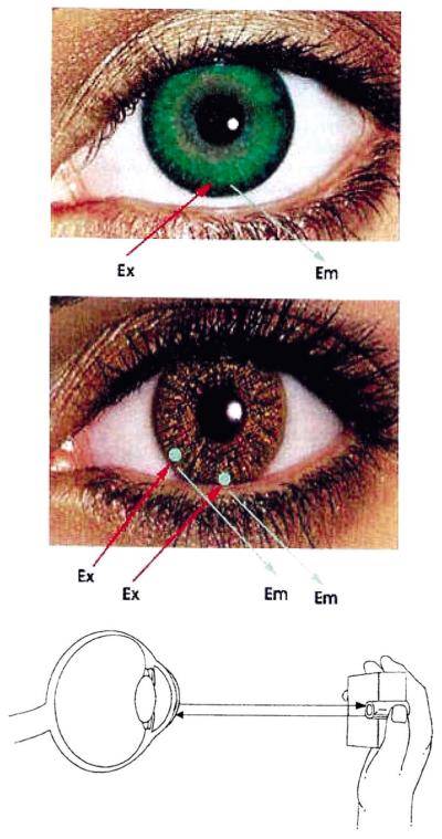
Fig. 2.
Potential methods for non-invasive continuous tear glucose monitoring. (Top) BAF doped contact lens as described here, and (Middle) Sensor spots on the surface of the lens to additionally monitor other analytes in addition to glucose, such as drugs, biological markers, Ca2+, K+, Na+, O2 and Cl−. Sensor regions may also allow for ratiometric, lifetime or polarization based fluorescence glucose sensing. (Bottom) Schematic representation of the possible tear glucose sensing device.

