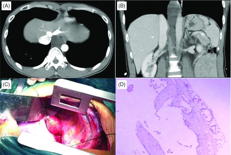
Fig. 1.
(A) CT scan showing an oval, homogeneous, non-enhancing cystic mass in the posterior mediastinum. (B) Coronal scan showing a large dilated cyst lesion from the level of right inferior pulmonary vein to the retroperitoneum. (C) Intraoperative findings. (D) The cyst wall consists of fibrous connective tissue and small foci of lymphoid cell aggregation (hematoxylin-eosin, original × 4).

