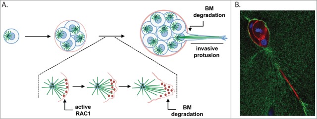
Figure 1.
Centrosome amplification induces the formation of invasive protrusions. (A) Schematic representation of mammary epithelial cells (MCF10A) with extra centrosomes growing in 3-D cultures. Single cells plated on top of a mix containing matrigel:collagen-I will divide and form small spheres, called acini. In cells with extra centrosomes, acini will form long, invasive protrusions that are able to degrade the basement membrane (BM). This process requires the activation of RAC1 downstream of microtubules in cells with extra centrosomes. We propose that localized activation of RAC1 is determined by the polarization of the microtubule cytoskeleton toward the cell cortex. After the initial activation of RAC1, changes in the cell cortex, such as Arp2/3-mediated actin polymerization and lamellipodia formation, will determine the formation of the invasive protrusion. (B) Invasive acini containing extra centrosomes. Cells were stained for F-actin (red), fibronectin (green) and DNA (blue). One predominant invasive protrusion can usually be observed in these acini. Increased accumulation of fibronectin surrounding the invasive protrusions can also be detected.

