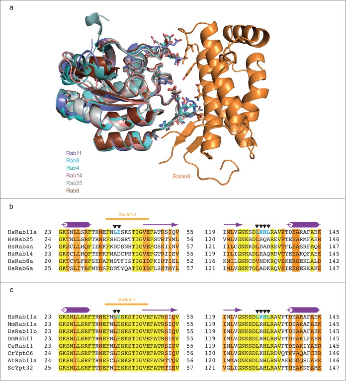
Figure 4.

The Rabin8-binding interface is unique to Rab11. (A) Structural overlay of different active Rab GTPases onto the Rab11-GMPPNP-Rabin8 crystal structure shown as a cartoon representation. Rab4 (PDB code: 1YU9), Rab6 (PDB code: 1YZQ), Rab8 (PDB code: 4LHW), Rab14 (PDB code: 4D0G) and Rab25 (PDB code: 3TSO) are cartooned in different colors. Residues utilized by Rab11 to bind Rabin8 and the corresponding residues in different Rab GTPases are shown as sticks. (B) Sequence alignment of Rabin8-interacting regions in human Rab11a with other human Rab GTPases. Rab11a residues that interact with Rabin8 are indicated with black arrows and labeled in bold blue color. Highly conserved residues are highlighted in yellow, similar residues in lighter and darker orange, respectively. Secondary structure derived from human Rab11a is indicated above the sequence. (C) Multiple sequence alignment of Rab11a protein from different species. Rab11a residues that interact with Rabin8 are indicated as shown in Fig 4B. Secondary structure elements from Rab11a structure are indicated above the sequence. Hs: Homo sapiens, Mm: Mus musculus, Dm: Drosophila melanogaster, Ce: Caenorhabditis elegans, Cr: Chlamydomonas reinhardtii, At: Arabidopsis thaliana, Sc: Saccharomyces cerevisiae.
