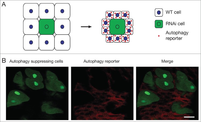
Figure 2.
Cell-autonomous function of autophagy genes. (A) Experimental design. A cell with Atg gene knockdown by RNAi co-expresses green fluorescent protein (GFP, green) and is surrounded by wild type cells (white). After autophagy induction, red autophagy reporter punctae are present only in wild type cells, and not in the Atg RNAi knockdown cell. The Atg RNAi knockdown cell is larger than wild type cells, because autophagy leads to a reduction in cell size. (B) An image of the mid gut of the Drosophila larval intestine. Cells with Atg1 knockdown are marked by GFP (green). mCherry-tagged Atg8a protein (red), which localizes to autophagosomes and autolysosomes, is expressed in all cells and serves as a reporter of autophagy. Two hours after puparium formation (APF), mCherry-Atg8a punctae are only present in the wild type mid gut cells, but not in the Atg1 RNAi-expressing green cells. Scale bar represents 50 μm. Image courtesy of T.-K. Chang.

