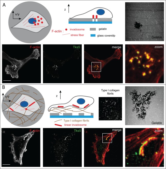
Figure 1.
Type I collagen induces linear invadosomes. (A) Schematic representation and confocal images of cancer cells seeded on fluorescent gelatin exhibiting classical invadosomes. Invadosomes are organized in individual dots and degrade the underlying extracellular matrix. Shown are representative confocal images of cells stained for tyrosine kinase substrate 5 (Tks5, also known as Fish; green) and filamentous actin (F-actin; red). Gelatin was stained with fluorescein (gray). Panels on the right show a 4× zoom of the white squares. Scale bar: 10 μm. (B) Schematic representation and confocal images of cancer cells seeded on type I collagen fibrils and fluorescent gelatin presenting as linear invadosomes. Linear invadosomes are aligned along fibrils and degrade gelatin and the collagen fibrils themselves. Shown are representative confocal images of cancer cells stained for Tks5 (green) and F-actin (red). Collagen I fibrils were labeled with 546-succinimidyl-ester (gray) and gelatin was stained with fluorescein (gray). Panels on the right show 4x zoom of the white squares. Scale bar: 10 μm.

