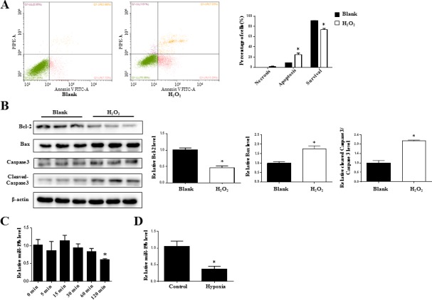
Figure 2. miR-19b is decreased in H2O2-treated H9C2 cardiomyocytes.

A. Flow cytometry analysis for necrosis, apoptosis, and survival of H9C2 cardiomyocytes treated with H2O2 (600 μM, 2 h) (n = 3). B. Immunoblot analysis for Bcl-2, Bax, cleaved-Caspase 3 to Caspase 3 ratio in H2O2-treated H9C2 cardiomyocytes (600 μM, 2 h) (n = 3). C. The time-course change of miR-19b as determined by qRT-PCRs in H2O2-treated H9C2 cardiomyocytes. (n = 5) D. The downregulation of miR-19b in hypoxia treatment. (n = 5) *, P < 0.05.
