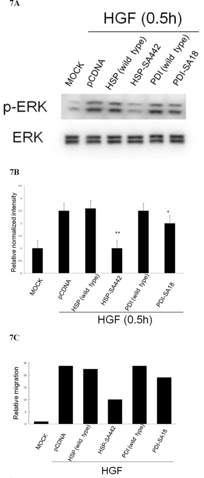
Figure 7. Alteration of cysteine on HSP60 prevented HGF-induced ERK phosphorylation and HepG2 cell migration.
HepG2 cells were transiently transfected with none (MOCK), pcDNA vector or wild type or cysteine mutant of HSP60 and PDI for 36 h followed by treatment with 25 nM HGF for 30 min (A) (B), or 48 h (C). Western blot of p-ERK (A) and transwell cell migration (C) were performed. In (A) ERK was used for normalizing the band intensities. (B) is the quantitative figures for (A). (**) represent statistical significance (Student's t test: p < 0.005, N = 3) (**) and (*) represent statistical significance (Student's t test: p < 0.005, and p < 0.05, respectively, N = 3) for differences of relative normalized intensity between HSP-SA442 (the HSP60 mutant) and wild type HSP60, and that between PDI-SA18 (the PDI mutant) and wild type PDI. In (C), relative migratory activity for each treatment was calculated as average of two reproducible result, taking the data of MOCK sample as 1.0.

