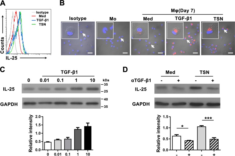
Figure 3. TGF-β1 promotes the production of IL-25 by Mφs in gastric cancer tissue.
(A) Flow cytometric and (B) immunofluorescent analyses show the expression levels of IL-25 in uncultured monocytes (Mo) and monocytes cultured in medium alone (Med), TGF-β1 (TGF-β1) or supernatant from an SGC7901 gastric cancer cell line (TSN). Scale bar = 30 μm. Negative controls were prepared using isotype-matched antibodies. (C) Western blotting shows the dose dependence of IL-25 expression in monocytes cultured with TGF-β1 at the indicated concentrations (ng/mL). (D) Western blotting shows the effect of blocking TGF-β1 in monocytes cultured with TSN: (−), untreated monocytes; (+), monocytes pretreated with a blocking antibody against TGF-β1. GAPDH was used as an internal control. Semi-quantitative evaluations were performed using Image J software; *p < 0.05; ***p < 0.001.

