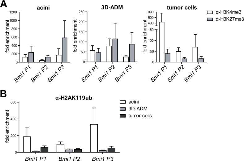
Figure 5. Histone modifications on the Bmi1 promoter were changed in the in vitro sequence of pancreatic carcinogenesis.
(A) Epigenetic signature of three Bmi1 promoter regions (P1 to P3 in the direction from 5′ to 3′) analyzed with ChIP-RT-PCR using H3K4me3 and H3K27me3 antibody represented in three panels for acini, 3D-ADM and tumor cells. (B) Direct comparison of the H2AK119ub loss on the Bmi1 promoter regions in the in vitro sequence of acini, 3D-ADM and tumor cells. All ChIP-RT-PCR data are calculated as fold enrichment over IgG control and are represented as mean ± SEM (n = 3–5).

