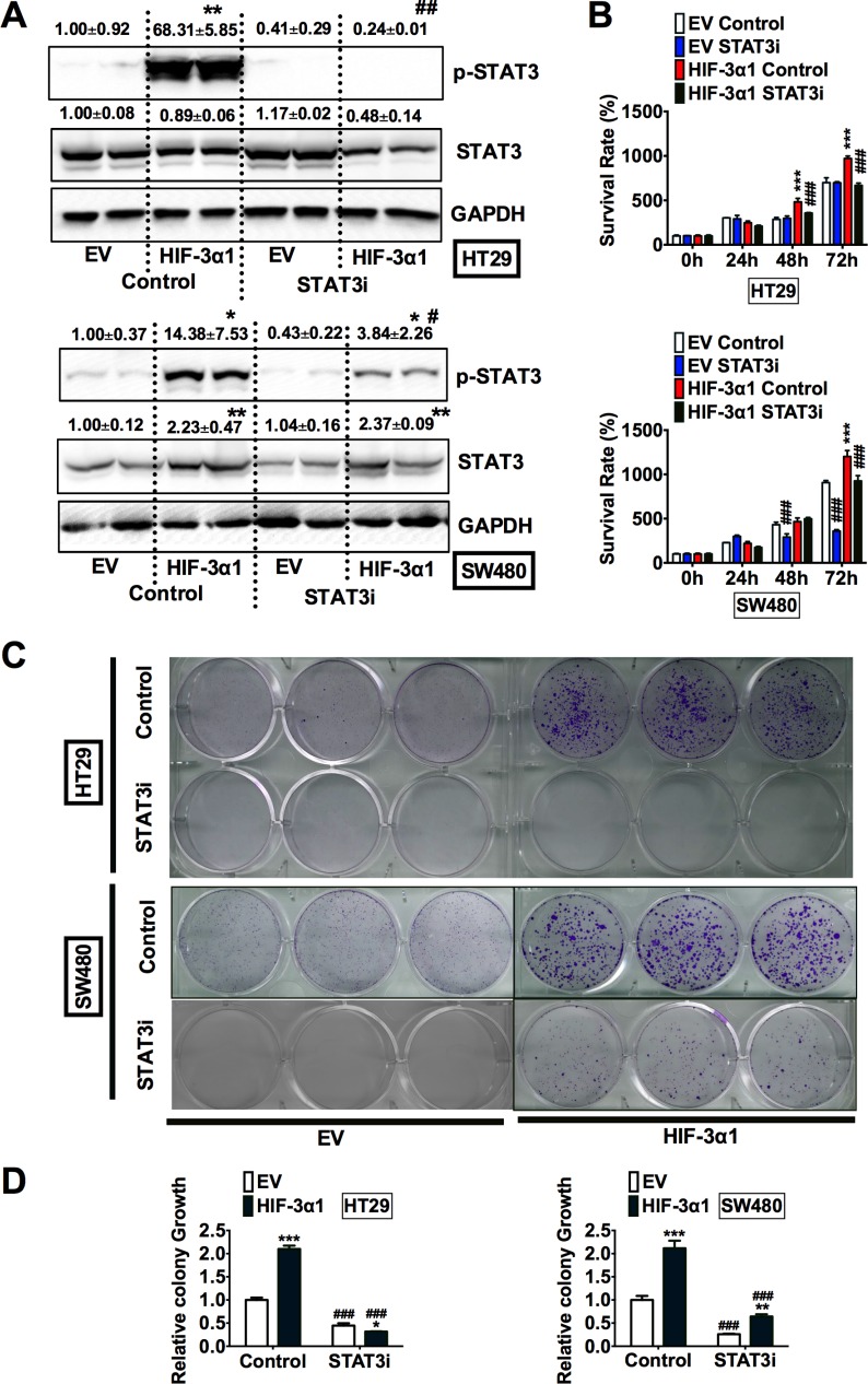
Figure 5. Inhibition of STAT3 decreases HIF-3α1-enhanced CRC cell and colony growth.
(A) Western blot analysis of p-STAT3 and STAT3 in whole cell extracts, (B) cell survival determined by MTT assay, (C) colony formation detected by crystal violet assay and (D) quantification of colonies formed from STAT3 inhibitor (STAT3i) treated or untreated HIF-3α1-overexpressing or EV lentivirus-infected HT29 or SW480 CRC cells. *p < 0.05, **p < 0.01, ***p < 0.001 compared with EV control cell line. #p < 0.05, ##p < 0.01, ###p < 0.001 compared with untreated controls.

