
Figure 5. Targeting hypoxia by cetuximab decrease TFE3 in Tgfbr1/Pten 2cKO mice.
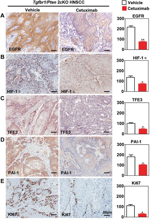
Immunohistochemical staining of HIF-1α, TFE3, PAI-1, EGFR and proliferating marker Ki67 in Tgfbr1/Pten conditional knock out mice after cetuximab treatment and with quantitative analysis. The expression of HIF-1α, TFE3, PAI-1, EGFR and Ki67 were was significantly reduced in the cetuximab treatment group than the vehicle group (t test, *, P<0.05, **, P<0.01). Scale bars =50μm.