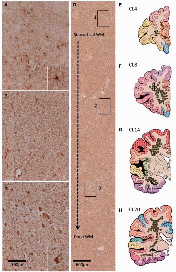
Figure 2.
Distribution of GFAP+ clasmatodendrocytes in the deep white matter regions of post-stroke survivors. ( A–C ) Panels show normal appearance of GFAP+ astrocytes in the immediate superficial layers of the white matter ( A ), retracted astrocytes at mid-level ( B ) and clasmatodendrocytes in the deep white matter, with a particularly high concentration at the level of the anterior horn of the lateral ventricles ( C ). Insets in A and C show a higher magnification of the different forms of GFAP+ astrocytes predominant in A and C , respectively. In C inset, cell vacuolation is evident. Boxes 1–3 in D delineate location of images ( A–C ), demonstrating the distribution of GFAP+ cells from the immediate subcortical layer to the deep white matter. Illustrative coronal sections ( E–H ) shows the distribution of clasmatodendrocyte densities (brown dots) in the white matter regions at different coronal levels incorporating the frontal, temporal and parietal lobes. Box in F represents approximate location of image D. Scale bars = 100 μm ( A–C ); 20 μm (insets). WM = white matter.

