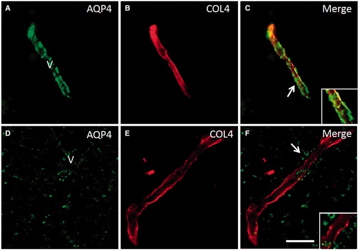
Figure 5.
Redistribution of AQP4 from COL4 labelled microvessels and capillaries in the deep white matter in PSD. ( A–C ) Panels show a AQP4 and COL4 labelled capillary in deep white matter of a PSND case demonstrating localization of AQP4 immunoreactivity in the vessel wall ( C ). ( D–F and insets) show lack of co-localization of AQP4 and COL4 in regions where clasmatodendrocytes were found. The disrupted distribution of AQP4 is evident in F (arrow). Scale bar = 25 µm.

