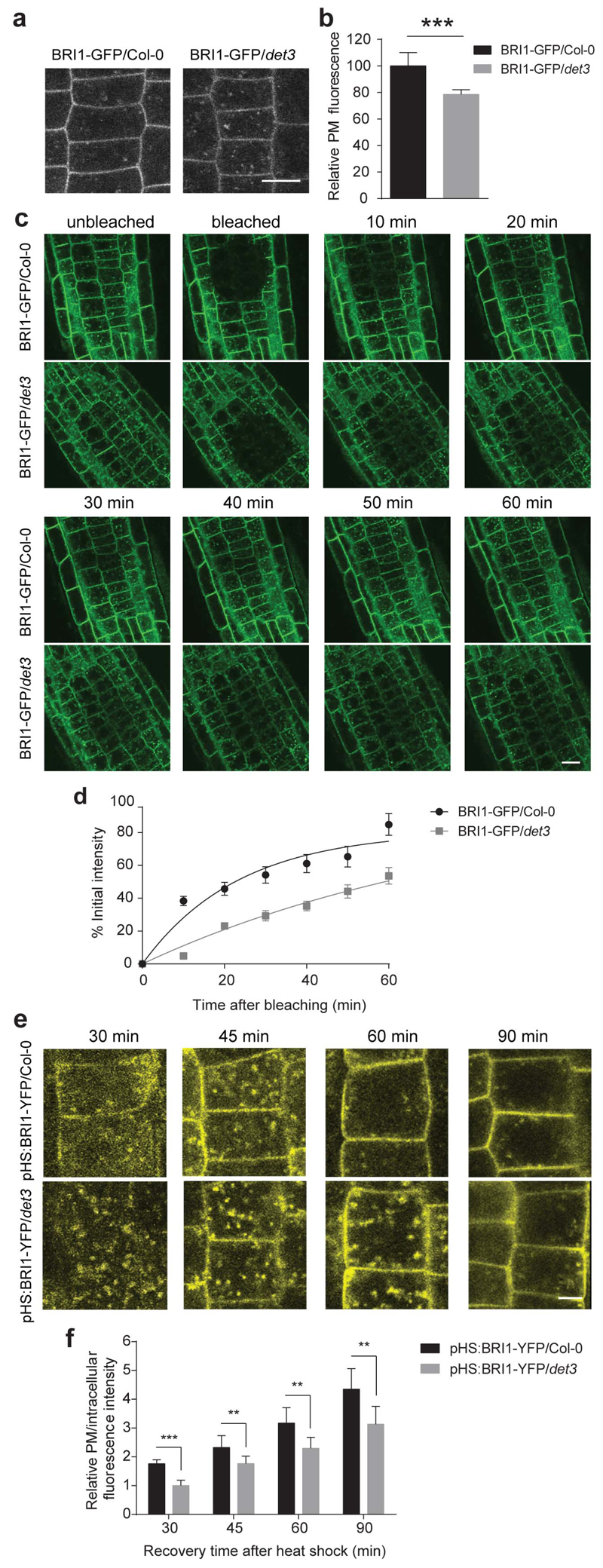
Figure 4. Secretion of BRI1 in det3 is reduced.
a, Confocal images of 5-day-old BRI1-GFP/Col-0 and BRI1-GFP/det3 root epidermal cells. Scale bar, 5 μm. b, Quantification of relative plasma membrane (PM) fluorescence intensity of BRI1-GFP/Col-0 and BRI1-GFP/det3 as shown in a (30 cells from five roots were measured). Plasma membrane fluorescence was normalized to background fluorescence for each measurement. P values (t -test), *** P< 0.001 relative to the respective control. c, FRAP analysis of BRI1-GFP/Col-0 and BRI1-GFP/det3 plasma membrane BRI1 recovery rate. Consistent ROI was selected for individual roots, followed by photobleaching to significantly reduce the target area fluorescence. Root epidermal cell fluorescence was recorded before bleaching and at different time points after bleaching. Scale bar, 10 μm. d, The fluorescence recovery was calculated on cells with totally bleached whole plasma membrane fluorescence. Plasma membrane fluorescence before and immediately after bleaching was set as 100 % and 0 %, respectively. Each recovery value was normalized to fluorescence value of the unbleached region (at least 15 cells from three roots were measured). e, In vivo analysis of heat shock-induced BRI1-YFP exocytosis. YFP signal in 5-day-old pHS:BRI1-YFP/Col-0 and pHS:BRI1-YFP/det3 root epidermal cells chased at 30 min, 45 min, 60 min, and 90 min after 1-h 37°C induction. Scale bar, 5 μm. f, Relative plasma membrane to intracellular fluorescence intensity values calculated by fluorescence intensities measured using ImageJ. ROI was kept constant for each measurement (at least 15 cells from three roots were measured). Error bars indicate S.D.

