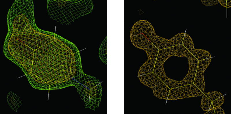
Figure 7.13.13.

Neutron versus X-ray macromolecular crystallography. Left: The neutron density for Tyr137 in the structure of D-xylose isomerase contoured at 1.5σ (green) and 2.0σ (yellow) clearly reveals the orientation of the deuteron on the O atom of tyrosine. Right: The protonation state of Tyr254 remains unclear from electron density maps in the X-ray crystal structure of the same enzyme determined to 0.94-Å resolution at −170°C. Reprinted from Hanson et al. (2004) with permission from the International Union of Crystallography.
