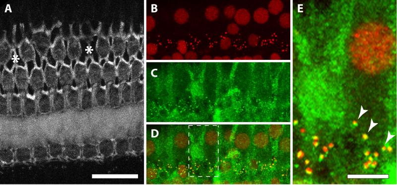
Figure 1.
Images from a cochlear segment below 1 kHz. A. Phalloidin-labeled cuticular plates of hair cells. Asterisks represent missing 3rd row OHCs. B. IHC nuclei and presynaptic ribbons both label with anti-CtBP2 (from the same cochlear segment as in A). C. Post-synaptic receptors and cytoplasmic background label with anti-GluR2. D. Merged images from B and C. Dashed lines indicate inset displayed in E. Scale bar in A is shared for A-D. E. Arrowheads show the co-localization of CtBP2 (red) and GluR2 (green).

