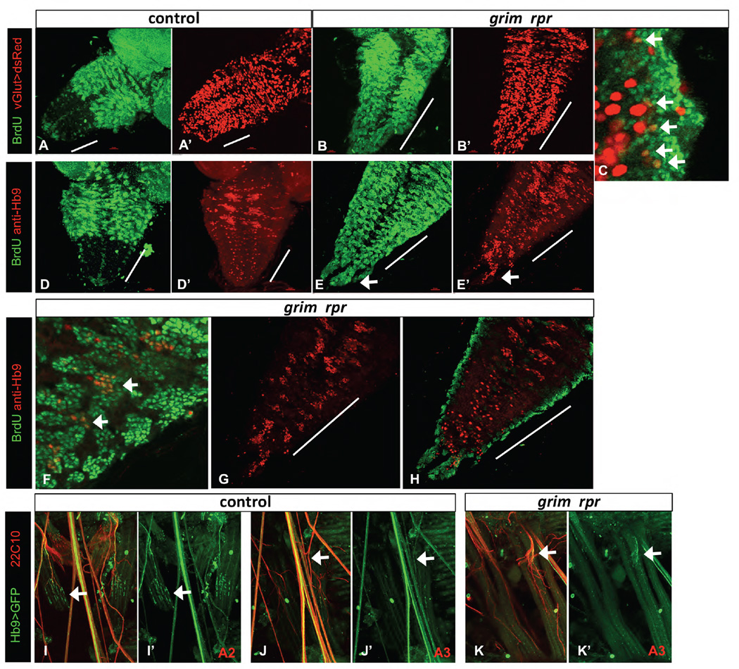
Figure 5. Ectopic vGlut and Hb9 expressing neurons are born post-embryonically in the grim rpr mutant.
BrdU feeding of larvae was used to label neurons born post-embryonically. The grim rpr mutant (3.5A/MM2) (B, C, E, F–H) has a dramatic increase in surviving and proliferating NB lineages in the abdominal segments (marked with a white bar). A–C) vGlut-Gal4 UAS-dsRed (red) marks glutamatergic motor neurons and some additional neurons. A–A’) Control larval VNC. B–C) There is an increase in BrdU positive (green) dsRed co-labeled cells in the abdominal segments of the grim rpr mutant (arrows in C) compared to control. D–H) Hb9 expressing neurons (red) are born post-embryonically in the thoracic segments of the wild type VNC and appear in three pairs of clusters (D), but not in the abdominal segments (abdominal segments marked with a white bar). In the grim rpr mutant, medial clusters of post-embryonically born Hb9 expressing cells are present in each abdominal segment (E, arrows in F), including in the terminal extensions seen in these mutants (arrows in E, E’). G shows Hb9 alone in double labeled ventrally located post-embryonically born clusters, while H shows increased embryonically born, more dorsal Hb9 positive cells. Both are increased in the grim rpr mutant. I–K’) Innervation of specific segments by Hb9 positive motorneurons. Strong ventral muscle innervation by Hb9 expressing motorneurons can be seen in the second abdominal segment (A2) of control animals (I–I’). However, this innervation is much reduced in the third segment (A3) of those same individuals (J–J’). Innervation by Hb9 expressing motorneurons in the grim rpr mutant is unchanged compared to control (K–K’ and J–J’).

