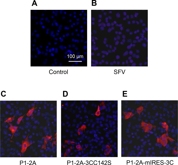
Fig 2. Expression of FMDV capsid proteins using the SFV split-helper system.
Uninfected BHK cells (A) or cells infected with rSFV (B), rSFV-FMDV-P1-2A (C), rSFV-FMDV-P1-2A-3CC142S (D) or rSFV-FMDV-P1-2A-mIRES-3C (E), at an MOI of 20, were immunostained at 16 h post infection. FMDV proteins were detected with an anti-FMDV O1 Manisa polyclonal antibody and a secondary antibody labeled with Alexa Fluor 568 (red). The cellular nuclei were visualized with DAPI (blue). Bar, 100 μm. The results shown are representative of three independent experiments.

