
Fig 3. Optical coherence tomography scan images (1:1 pixel views) showing the representative images in eyes with unilateral traumatic optic neuropathy (TON).
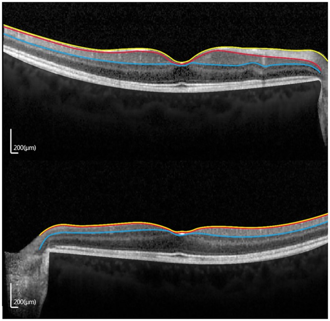
(A) Contralateral normal eye. (B) TON eye. (RNFL, retinal nerve fiber layer; GCIPL, ganglion cell layer and inner plexiform layer.).