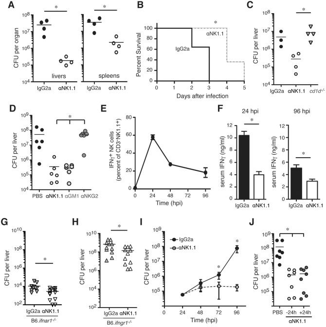
Fig 1. NK cells increase susceptibility to systemic L. monocytogenes (Lm) infection despite an independent from early IFNγ production.
(A) Lm burdens from C57BL/6 (B6) mice treated with control (IgG2a) or NK1.1+ cell depleting (αNK1.1) antibodies (Abs) and infected 24 h later with 104 Lm. Shown are total colony forming units (CFU) per tissue as determined by dilution plating of tissue lysates at 96 h post-infection (hpi). Symbols represent individual mice with mean (lines). Shown is one of five experiments using n = 3–5 mice/group. (B) Survival curve for B6 mice treated with IgG2a or αNK1.1 antibodies and infected 24 h later with 5 x 10⁴ Lm. Data are pooled from two experiments using n = 5–7 mice/group (C) Lm burdens from livers of B6 and NKT cell deficient (B6.CD1d -/-) mice at 96 hpi. B6 mice were treated with IgG2a or αNK1.1 24 h before infection. Liver CFU from one of three experiments using n = 3–4 mice/group. (D) Lm burdens from livers of B6 mice treated 24 h before infection with αNK1.1+, anti-asialoGM1 (αGM1), or a non-depleting anti-NKG2A/C/E (αNKG2) Ab. Liver CFU at 96 hpi are shown for two pooled experiments representing n = 6 mice/group. (E) Proportions and total cell numbers of splenic NK1.1+CD3- gated NK cells staining positive for intracellular IFNγ at the indicated hpi. Shown are mean ± SEM for data pooled from 3–5 experiments representing n = 9–18 mice/time point. (F) Serum IFNγ measured for control IgG2a or αNK1.1 treated B6 mice at 24 or 96 hpi. (G) B6.ifnar1 -/- mice were treated with indicated Abs 24h before infection as above. Shown are liver bacterial burdens at 96 hpi. Bars depict mean ± SEM values from 3 pooled experiments using n = 3–5 mice/group. (H) B6.ifngr1 -/- mice were treated with indicated Abs 24h before infection with 4 x 10³ CFU Lm. Livers were harvested for CFU counts at 72 hpi. Bars depict mean ± SEM values from 3 poled experiments using n = 3–5 mice/group. (I) B6 mice were treated with antibodies and infected as in (A). Liver bacterial burdens at 24, 48, 72, and 96 h after Lm infection. Data are mean ± SEM from 3–5 pooled experiments representing n = 9–18 mice/time point. (J) B6 mice were left untreated or administered αNK1.1 at 24 h before or 24 h after Lm infection. Data are pooled from two experiments with n = 3–4 mice/group. *P<0.05 as measured by (A, F-H) t-test or (B, D, I, J) ANOVA.

