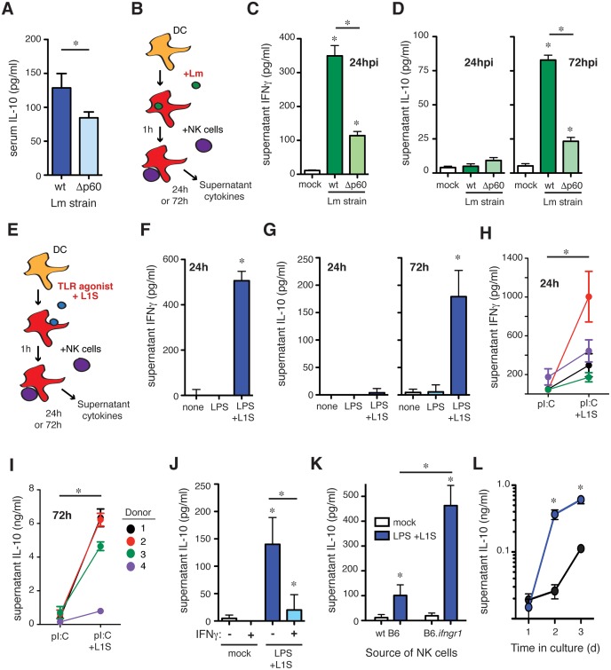
Fig 3. The L1S region of the Lm p60 protein is sufficient to induce NK cell IL-10 production.
(A) Serum IL-10 at 96 hpi in B6 mice infected with wt or p60-deficient (Δp60) Lm strains. (B) Schematic of co-culture system consisting of BMDC infected with Lm for one hour, followed by washing and the addition of splenic purified NK cells at 2 h post-infection. (C) 24 h supernatant IFNγ after infection of BMDC with wt or p60-deficient Lm in co-culture with wt NK cells. (D) 24 h and 72 h supernatant IL-10 after infection of IL-10 deficient BMDC with wt or p60-deficient Lm in co-culture with wt NK cells. (E) Schematic of co-culture system consisting of BMDC treated with a TLR agonist ± L1S for one hour, followed by washing and the addition of splenic purified NK cells at 2 h post-stimulation. (F) 24 h supernatant IFNγ after treatment of BMDC with LPS ± L1S in co-culture with wt NK cells. (G) 24 h and 72 h supernatant IL-10 after treatment of IL-10 deficient BMDC with LPS ± L1S in co-culture with wt NK cells. (H) 24 h supernatant IFNγ after treatment of human DC with polyI:C ± L1S in co-culture with PBMC from unrelated donors. (I) 72 h supernatant IL-10 after treatment of human DC with polyI:C ± L1S in co-culture with PBMC from unrelated donors. (J) 72 h supernatant IL-10 after treatment of IL-10 deficient BMDC with LPS + L1S ± stimulation with 50 ng/mL IFNγ in co-culture with wt NK cells. (K) 72 h supernatant IL-10 after stimulation of IL-10 deficient BMDC with LPS + L1S in co-culture with NK cells from wt or IFNGR-/- mice. (L) IL-10 was measured in supernatants collected at 24, 48, and 72 h from L1S-stimuated co-cultures of IL-10 deficient BMDC and NK cells from wt or IFNGR-/- mice. Data for (C, D, F, G, H, I, J, K, L) pooled from three or more experiments. *P<0.05 as determined by t-test.

