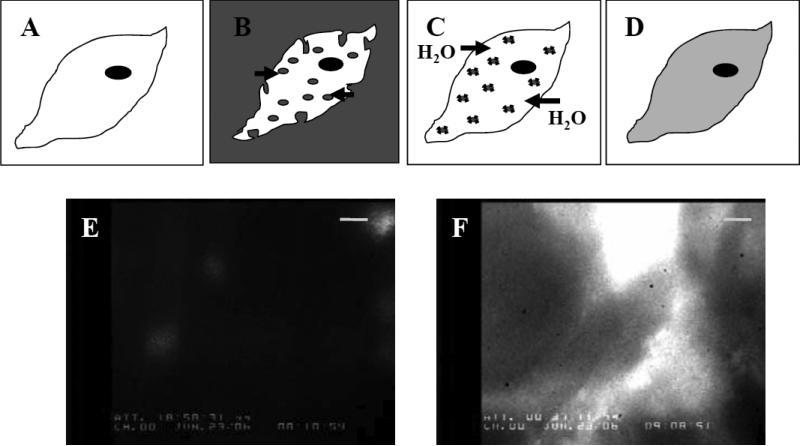
Fig. 1.
Hypertonic loading of chick cardiomyocytes in culture. Panels (A)–(D) are a cartoon representation of the hypertonic loading protocol used to introduce nucleotide into chick cardiomyocytes. A: Chick myocyte in isotonic plating media. B: Hypertonic loading media (see Materials and Methods) containing total 6 mM nucleotide (either ATP, dATP, or a combination of the two) applied to the cell; note that in some instances, loading media also contained a fluorophore (Materials and Methods). Pinocytotic vesicles carry hypertonic loading media into the cell as water is released from the cell to the hypertonic media. C: Hypotonic lysis media applied to the cell. Pinocytotic vesicles lyse within the cell cytoplasm as water enters the cell, thus releasing nucleotide. D: Cell recovery in isotonic plating medium. Panels (E) and (F) demonstrate fluorescein as an indicator of successful hypertonic loading, using fluorescence microscopy with a FITC filter at 100x magnification. Calibration bar represents 10 μm. Panel (F) shows a different region of the same cultured monolayer as panel (E). Panel (E) corresponds to (A), and panel (F) corresponds to (D). E: Chick cardiomyocyte monolayer prior to hypertonic loading. F: Monolayer after hypertonic loading with 20 μM fluorescein in the hypertonic loading media (B), and following pinocytotic lysis (C) and recovery (D). Loading of applied extracellular solution is validated by fluorescein incorporation into the cells during treatment.

