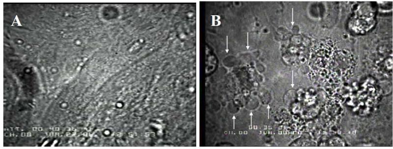
Fig. 4.
Changes in cell structure induced when intracellular [dATP] was increased to ≥100 μM. A: Representative confluent beating monolayer of embryonic chick cardiomyocytes prior to hypertonic loading. B: Changes in cell structure consistent with apoptosis following treatment to increase the intracellular [dATP] to ≥100 μM (Fig. 2). Cells are no longer confluent, and are rounded with membrane blebbing (arrows). Blebbing was observed in 100% of the monolayers treated with 6 mM hypertonic loading solution, but never at lower levels of dATP loading.

