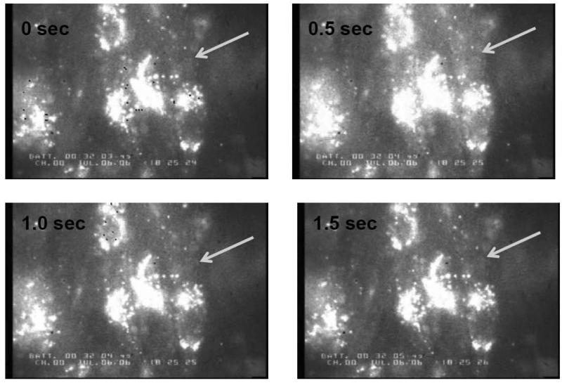
Fig. 6.
Recording of cytoplasmic Ca2+ in cultured chick cardiomyocytes using Rhod-2 AM calcium indicator. Changes in cytoplasmic Ca2+ were tracked over time by observing the increase in cytoplasmic fluorescence from time 0 to the peak of the Ca2+ transient (0.5 s), and decrease back to baseline. Arrow points to cytoplasmic region of interest (ROI). ROI's were chosen to avoid Rhod-2 accumulation within organelles, which appear as bright spots within the cell monolayer (Materials and Methods). Representative images are from ATP control following hypertonic loading at 25°C.

