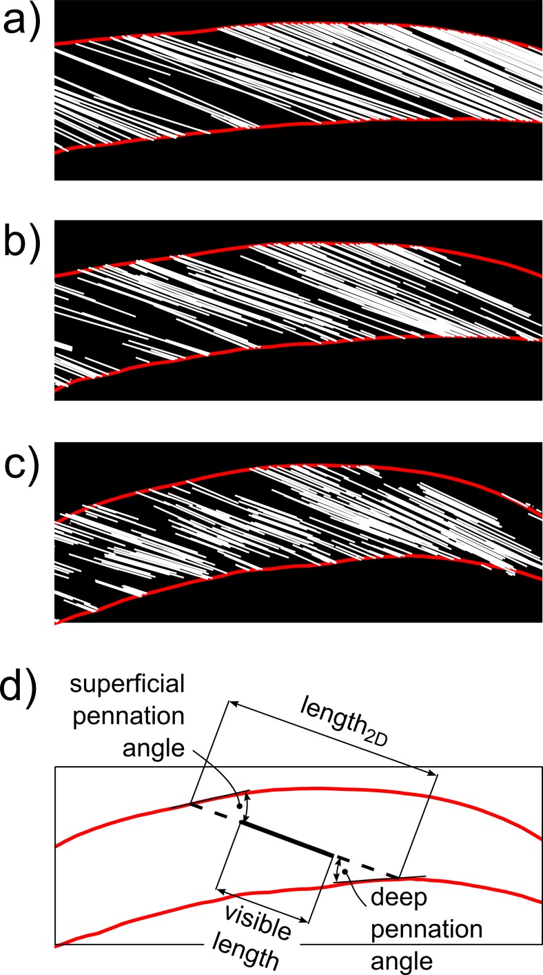
Fig 2. Examples of virtual ultrasound images.
Virtual ultrasound images of the medial gastrocnemius obtained with the transducer held at the same site on the skin, but under three different orientations which result in (a) good alignment between image plane and fascicles (misalignment is 1.3°), (b) reasonable alignment (7.5°) and (c) poor alignment (18.3°). Note that the fascicle sections become shorter with increasing misalignment. (d) Same virtual ultrasound image as shown in (c) but with only one fascicle section (solid black line) and its 2D reconstruction (dashed black line), obtained by extending the fascicle section to its intersections with the aponeuroses. For each fascicle section, the length and pennation angle in the 2D image plane were calculated as shown.

