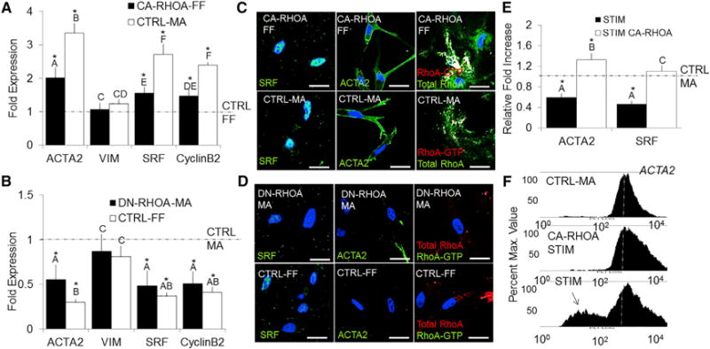
Figure 3. Myofibroblastic Activation in HH40 AV Progenitor Cells Is Dependent on Active RhoA.

(A and B) HH40 cells were isolated from left AV tissue and immediately transfected with CA-RhoA or DN-RhoA (Neon Transfection system). mRNA expression for ACTA2, VIM, SRF, and CyclinB2 was quantified via real-time PCR and compared to CTRL-MA or CTRL-FF conditions.
(C and D) Immunofluorescence of SRF (green, left column), ACTA2 (green, middle column), and RhoA-GTP/RhoA (green/red, right column) in the collagen gels for each conditions.
(E) Transfected CA-RhoA cells were stretched (STIM) for 48 hr at 2.1 Hz, and mRNA expression of ACTA2 and SRF was quantified using real-time PCR.
(F) Flow cytometry was used to quantify ACTA2 protein expression in CTRL-MA, STIM, and CA-RhoA-STIM conditions (10,000 cells).
All samples were counterstained for nuclei using Draq5 (blue). Scale bars represent 10 μm (C and D). Error bars show ± SEM; n ≥ 4. One-way ANOVA with Tukey’s post hoc was performed. Different letters indicate statistical significance p < 0.05 from each other, and asterisks indicate significance from control. See also Figure S3.
