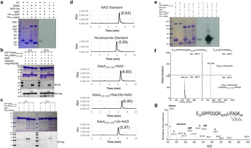
Extended Data Figure 10. Detection of the ubiquitination intermediate by using SdeA519-1100.
a, Full-length SdeA cannot produce 32P-labeled product in reactions using 32P-α-NAD. Reaction samples resolved by SDS-PAGE were detected by Coomassie staining (left panel) and then by autoradiography (right panel). Note the 32P-α-AMP-GST-ubiquitin complex can be detected in the reaction containing E1 but not SdeA. b-c, SdeA519-1100 is defective in auto-ubiquitination. Reactions containing the indicated components were allowed to proceed for the indicated time duration and the production of ubiquitinated Rab33b (b) or SdeA519-1100 was detected by immunoblotting. d, SdeA519-1100 induces the production of nicotinamide from NAD and ubiquitin. Retention time for nicotinamide and NAD was first determined by HPLC and nicotinamide can only be detected in the reaction containing SdeA519-1100, NAD and ubiquitin. e, SdeA519-1100 induces the production of 32P-ADPR-labeled ubiquitin. GST-ubiquitin or GST-ubiquitinR42A was incubated with 32P-α-NAD and SdeA519-1100 for 6 h. Classical E1 incubated with GST-ubiquitin was included as a control. Samples resolved by SDS-PAGE before autoradiography (20 min) (right panel). Note that GST-ubiquitinR42A cannot be labeled by 32P. Data in panels a to e are one representative from two independent experiments with similar results. f, The detection of a peptide with m/z 737.33 corresponding to the tryptic peptide -E34GIPPDQQRLIFAGK48- containing one ADP-ribosylation site was detected only after ubiquitin was incubated with SdeA519-1100. As a loading control, another unmodified ubiquitin peptide -T55LSDYNIQK63- was detected in both control and treated samples. g, Tandem mass analysis revealed that ADP-ribosylation occurred on Arg42 evidenced by the extensive fragmentation of the ADP-ribosylation into adenine, adenosine, AMP and ADP ions. Although not as extensive, the fragmentation of the peptide backbone helps confirm the peptide sequence. Data shown in all panels are one representative from two independent experiments with similar results. a, b, c, e, Uncropped blots and autoradiograph images are shown in Supplementary Fig. 1.

