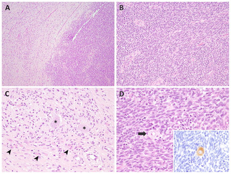
Figure 1. Microscopic features of MEM.
The most common appearance consisted of discrete sharply demarcated RMS (right) and ganglioneuroma (left) components (A, MEM2). The RMS component exhibited interlacing bundles of monomorphic spindle cells (B), while the ganglioneuroma component showed ganglion cells (asterix), nerve bundles, schwannian cells, and intermixed rhabdomyoblasts (arrowheads) (C, MEM2). Few ganglion cells (arrow) were scattered within the mitotically active RMS area (inset: synaptophysin stain) (D).

