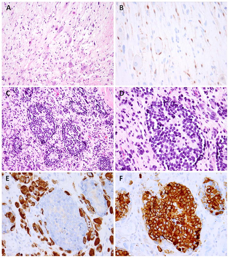
Figure 3.
Post-treatment MEM showed decreased cellularity and scattered rhabdomyoblasts and ganglion cells in a myxoid background (A, MEM4); an S100 stain highlights Schwannian and satellite cells (B). Another post-chemo MEM showed no treatment response, being composed of neuroblastoma islands with a micronodular pattern, within the RMS areas (C, MEM5). The neuroblastic cells have monomorphic round, hyperchromatic nuclei and form Homer-Wright rosettes (D). The desmin stain highlights the rhabdomyoblasts but not the neuroblastoma nodules (E), while synaptophysin stains the neuroblastoma nodules but not the surrounding rhabdomyoblasts (F).

