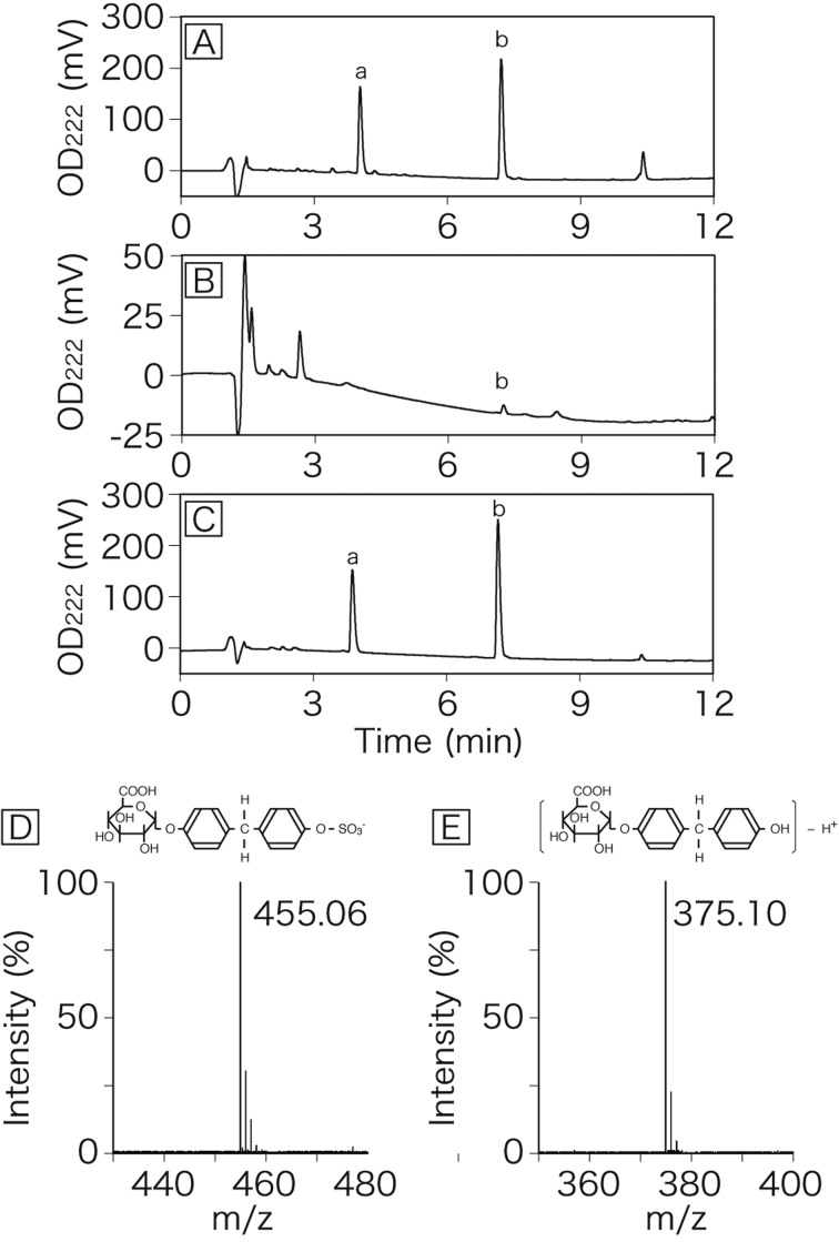
Fig. 1.
HPLC chromatograms of bile and venous perfusate derived from rat liver perfused with BPA. The liver was perfused for 5 min with Krebs-Ringer buffer containing BPA and then perfused with normal Krebs-Ringer buffer. Bile (A) and venous perfusate (B) were collected at 15 min after application of BPA to the liver. Peaks of BPA glucuronide/sulfate diconjugate (a) and BPA mono-glucuronide (b) are indicated. Panel C is a HPLC chromatogram of a standard solution of BPA conjugates. Panels D and E are a mass spectrum of peak a and peak b, respectively.

