
Figure 6.
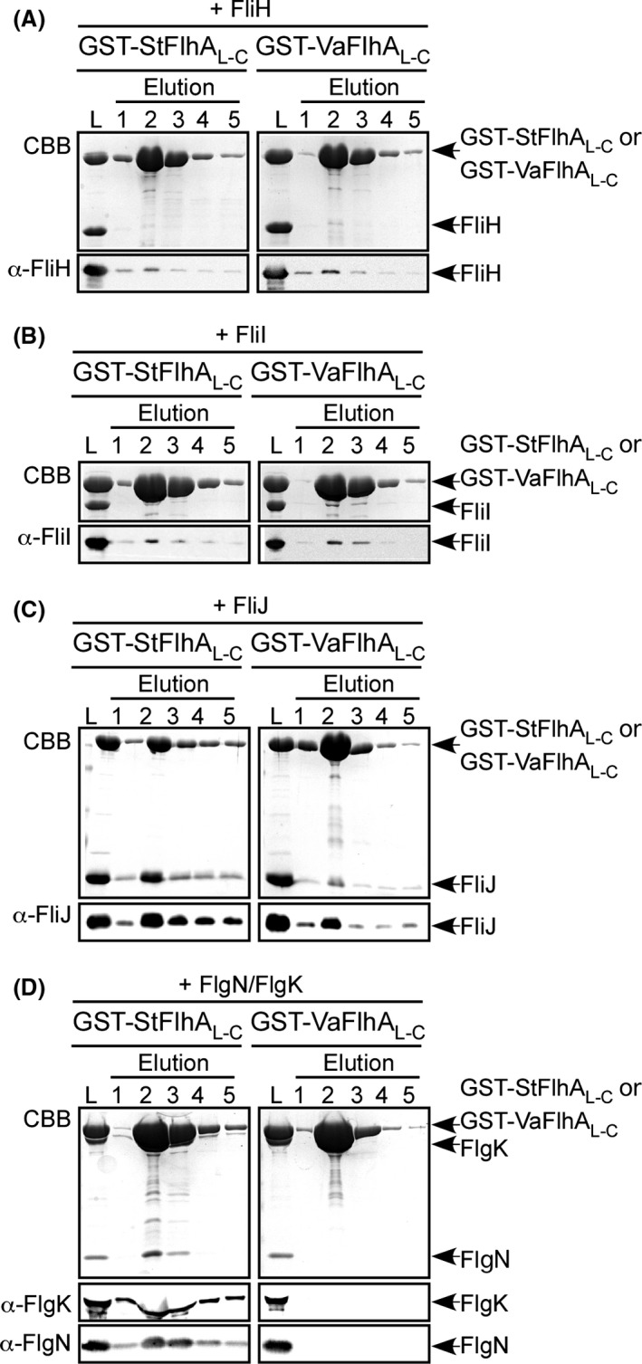
Interaction of Vibrio FlhA with FliH, FliI, FliJ, and the flagellar chaperone–substrate complex. Purified (A) FliH, (B) FliI, (C) FliJ, or (D) FlgN/FlgK complex was mixed with purified GST‐StFlhAL ‐C (left panel) or GST‐VaFlhAL ‐C (right panel), and dialyzed overnight against PBS. These mixtures (L) were loaded onto a GST column. After washing with 10 mL PBS, proteins were eluted with 10 mmol/L reduced glutathione. Elution fractions were analyzed by Coomassie Brilliant blue (CBB) staining (first rows) and immunoblotting by polyclonal anti‐FliH (A, second rows), anti‐FliI (B, second rows), anti‐FliJ (C, second rows), anti‐FlgK (D, second rows) or anti‐FlgN antibody (D, third rows).