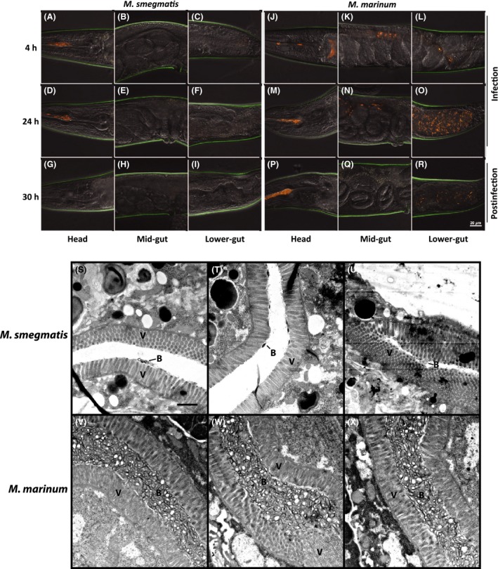
Figure 4.

Confocal and transmission electron microscopy of infected C. elegans. C. elegans infected with (A–C, G–I, M–O, S–U) M. smegmatis (tdTomato) or (D–F, J–L, P–R, V–X) M. marinum (tdTomato) were imaged at (A–F) 4 and (G–L) 24 h during infection and (M–X) 6 h postinfection (30 h). (A–R) The head, mid‐gut, and lower‐gut of 13–15 nematodes each at 4, 24, and 30 h were imaged using a confocal microscope and (S–X) at 30 h using transmission electron microscopy. A spectral filter for excitation wavelengths of 500–640 nm was used for confocal microscopy. The scale bar in R (20 μm) applies to panels A–R. The scale bar in S (1 μm) applies to panels S–X. V indicates nematode villi and B indicate mycobacterial debris and mycobacteria.
