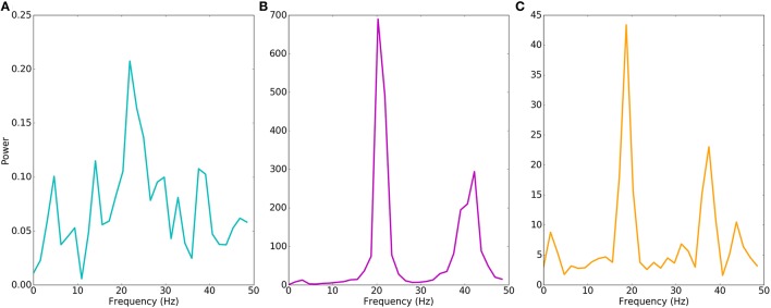
Figure 4.
Motor cortex models produce different beta oscillations. Power spectrum of multiunit activity vectors of examples in Figure 3. Power (y-axis) in arbitrary units. (A) Physiological model shows weak beta (22 Hz) oscillations with power of < 0.1% of the pathological model. (B) Pathological model produces strong beta (20 Hz) oscillations with additional harmonic at 40 Hz. (C) Epileptiform model produces strong beta (19 Hz) oscillations with additional harmonic at 38 Hz.

