Figure 1.
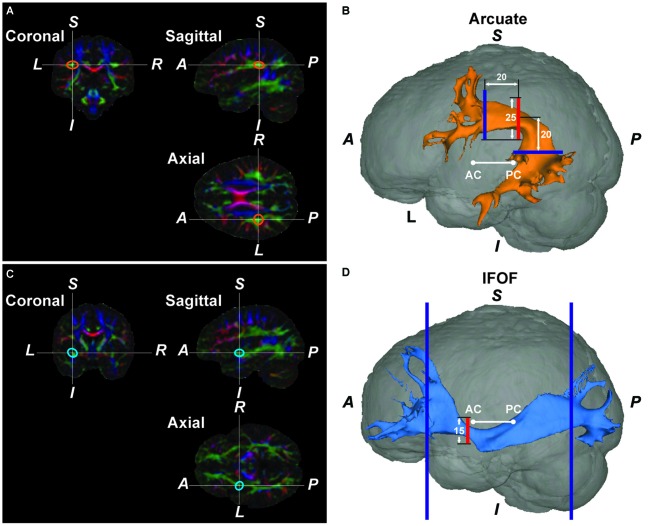
The Arcuate and inferior fronto-occipital fasciculus (IFOF) reconstructed by diffusion magnetic resonance imaging (MRI) in a typical participant. (A) A color-coded map of fiber bundles in the native space of a participant. In the Montreal Neurological Institute (MNI) space, we placed a “bottleneck mask” (a sphere of 10-mm radius) at a region where a narrower portion of the pathway was expected to pass. The mask was then transformed back to the native space. The orange ellipses denote the approximate outline of the transformed bottleneck mask on the left Arcuate. (B) The reconstructed left Arcuate. On a coronal slice, we generated a seed mask (shown in red), whose center was placed at the center of the transformed bottleneck mask on the left Arcuate. The waypoint masks are shown in blue. (C) A color-coded map of fiber bundles for the same participant. The blue ellipses denote the outline of the transformed bottleneck mask on the left IFOF. (D) The reconstructed left IFOF. On a coronal slice, we generated a seed mask (shown in red), whose center was placed at the center of the transformed bottleneck mask on the left IFOF. A, anterior; P, posterior; S, superior; I, inferior; L, left; R, right; AC, anterior commissure; PC, posterior commissure.

