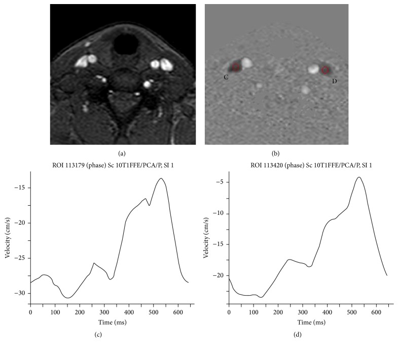
Figure 3.
2D Cine phase contrast at C5-C6 level: (a) magnitude image that displays carotids and IJVs; (b) phase image at the same level displaying dark flow directed to the heart and bright flow directed to the brain, with C and D indicating ROIs for flow measurements within IJVs bilaterally; and (c, d) velocity curves, respectively, for C and D ROIs.

