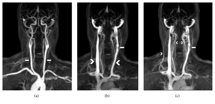
Figure 4.
Time-resolved MRV (4D-TRAK), reconstructed as coronal MIP projections at different time points, showing (a) arterial phase with carotid axes (arrows), (b) venous phase with visualization of IJVs (arrow heads), with a segmentary stenosis of left IJV (arrow), and (c) a delayed venous stage with better visualization of venous drainage of right external jugular vein and vertebral veins (arrow heads) and a confirmation of the left IJV stenosis from the previous phase (arrow).

