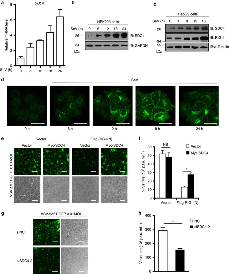
Figure 4. SDC4 is induced by virus infection and involved in regulating viral replication.
(a) HEK293 cells were infected with SeV for 6, 12, 18 or 24 h, followed by measurements of Sdc4 mRNA by quantitative real-time PCR analysis. (b,c) HEK293 cells (b) and HepG2 cells (c) were infected with SeV for different time courses, and analysed by immunoblotting. (d) HeLa cells were infected with SeV for 6, 12, 18 or 24 h, and then fixed with 4% paraformaldehyde, stained with an anti-SDC4 antibody, and observed by confocal microscopy. Scale bar, 50 μM. (e,f) HEK293 cells were transfected with the indicated expression vectors. Twenty-four hours later, the cells were infected with VSVΔM51-GFP at a MOI of 0.01 for 12 h. Subsequently, the cells were imaged by fluorescence microscopy (e), scale bar, 200 μM, or the culture supernatants were collected to measure the virus titre by a plaque assay (f). (g,h) HEK293 cells were transfected with siNC or siSDC4-2. Forty-eight hours after transfection, the cells were infected with VSVΔM51-GFP at a MOI of 0.01 for 12 h. Subsequently, the cells were imaged by fluorescence microscopy (g), scale bar, 200 μM, or the culture supernatants were collected to measure the virus titre by a plaque assay (h). The data in a,f and h are from one representative experiment of at least three independent experiments (mean±s.d. of triplicate assays). *P<0.05; NS, not significant versus the control groups.

