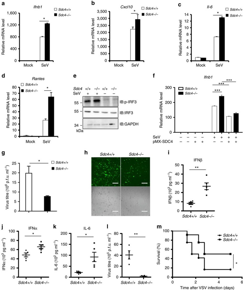
Figure 5. Enhanced type I IFN response in Sdc4-deficient cells.
(a–d) Primary Sdc4+/+ and Sdc4–/– MEFs were infected with SeV for 6 h, and analysed by quantitative real-time PCR (qRT-PCR). The transcript levels of Ifnb1 (a), Cxcl10 (b), Il6 (c) and Rantes (d) were measured by qRT-PCR analysis. (e) Primary Sdc4+/+ and Sdc4−/− MEFs were left untreated or infected with SeV for 6 h. The cell lysates were separated by SDS–PAGE and analysed by immunoblotting with the indicated antibodies. (f) Primary Sdc4+/+ and Sdc4−/− MEFs were first infected with a retrovirus expressing SDC4 or an empty vector. After 48 h of infection, the cells were infected with SeV for 7 h, and analysed by qRT-PCR. (g,h) Primary Sdc4+/+ and Sdc4−/− MEFs were infected with VSVΔM51-GFP at a MOI of 0.01 for 22 h. Subsequently, the culture supernatants were collected to measure the virus titre by a plaque assay (g) or the cells were imaged by fluorescence microscopy (h), scale bar, 200 μM. (i–k) Sdc4+/+ and Sdc4−/− mice (n=5 each) were infected with VSVΔM51-GFP via tail vein injection at 2 × 10⁸ p.f.u. per mouse. Sera were collected after 6 h infection to measure the levels of IFN-β (i), IFN-α (j) and IL-6 (k) by ELISA. (l) Sdc4+/+ and Sdc4−/− mice (n=5 each) were infected with VSVΔM51-GFP via tail vein injection at 1 × 10⁹ p.f.u. per mouse. Sera were collected after 12 h infection to measure the virus titre by a plaque assay. (m) Sdc4+/+ and Sdc4−/− mice (n=14 each) were infected with VSV virus via tail vein injection at 4 × 10⁸ p.f.u. per mouse and the survival of mice were monitored for 1 week. The data in a–d, f–g and i–l are from one representative experiment of at least three independent experiments (mean±s.d. of triplicate experiments in a–d and f, duplicate experiments in g). A two-tailed Student's t-test or log-rank (Mantel–Cox) test were used to analyse statistical significance. *P<0.05; **P<0.01; ***P<0.001 versus the control groups.

