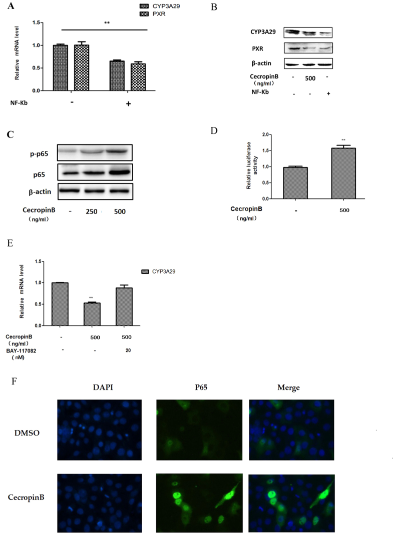
Figure 4. NF-κB activation is induced by cecropin B.
(A) HepLi cells were transfected with the NF-κB p65 over expression vectoror control vector for 24 hours, and CYP3A29/PXR mRNA level were detected by qRT-PCR. (B) HepLi cells were treated with 500 ng/ml of cecropin B or transfected with the NF-κB p65 over expression vector, and CYP3A29/PXR protein levels were detected by western blot. (C) HepLi cells were treated with 250 and 500 ng/ml of cecropin B for 12 hours, and NF-κB p65 and NF-κB p-p65 protein levels were detected by western blot. (D) HepLi cells were transfected with the NF-κB reporter plasmid, and cells were treated with or without 500 ng/ml of cecropin B after 24 hours. Luciferase activity was assayed at 12 hours after the treatments. E: HepLi cells were cotreated with 500 ng/ml of cecropin B and 20 nM of BAY117082 or 500 ng/ml cecropin B alone for 12 hours. The control group was treated with PBS. F: HepLi cells were transfected with the NF-κB p65-GFP overexpression vector for 24 hours, treated with 500 ng/ml of cecropin B for one hour, and fluorescence was examined under an OLMLPUS IX51 inverted fluorescence microscope. ** and * P < 0.01 and P < 0.05, respectively. The blot was cropped and the full-length blot is presented in Supplementary Fig. S2 (Fig. 4B,C).

