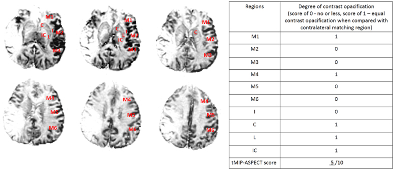
Figure 2. Six consecutive slices on PWI tMIP images that cover almost the whole territory of the middle cerebral artery were used to assess the extent of collateral filling.
tMIP-ASPECT score is based on scoring the degree of contrast opacification (0 - no or less, 1 – equal contrast opacification compared with matching region in contralateral hemisphere) in 10 regions, including C caudate, L lentiform nucleus, IC internal capsule, I insular ribbon and M1–6 cortical regions of the middle cerebral artery (M1–3 at the level of basal ganglia, M4–6 at the level rostral to the ganglionic structures). In this patient, the deficit of collateral blood flow involved M2–3, M5–6, and I, and the tMIP-ASPECT score was 5.

