Figure 4.

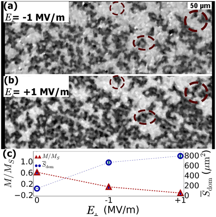
Kerr microscopy images of domain expansion under a modest out-of-plane magnetic field (−2.4 mT) and subsequent applications of electric fields to the PZT substrate of (a) −1 MV/m and (b) +1 MV/m (Circles in (a,b) highlight newly reversed areas due to electric field polarity switch). Changes in the magnetization (red triangles) and the average reversed (dark contrast) domain size (open blue circles) are shown in (c) under the series of applied electric fields.
