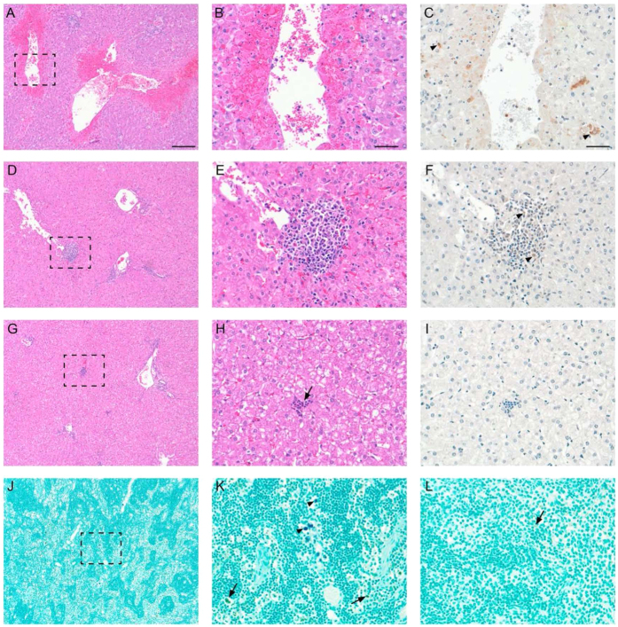
Figure 5. Liver and mesenteric lymph node histopathology and immunohistochemistry.
Rift Valley fever virus caused multifocal mid-zonal to central hepatic necrosis accompanied by neutrophilic and histiocytic inflammation. Additionally, hemorrhage was common in the larger necrosis lesions as seen in the 4 dpc mock-vaccinated, virus only study animals. (A,B) low and high power fields from a hematoxylin and eosin (H&E) stained section of liver parenchyma from a 4 dpc mock-vaccinated, virus only, sheep, #63, which had severe multifocal necrosis accompanied by hemorrhage involving ~15% of its hepatic parenchyma, hepatic histopathology score of 3. Each broken line box outlines the region shown at higher magnification in the next image. (C) Rift Valley fever virus antigen IHC on serial section of same tissue at high power, black arrowheads denote positive labeling for RVFV antigen, red-brown cytoplasmic signal in hepatocytes, inflammatory cells and cellular debris. (D,E) H&E stained liver section from a 7dpc mock-vaccinated sheep, #71, which had multifocal, 1–2 mm areas of necrosis with a more lymphohistiocytic infiltrate than #63’s liver and less than 5% of the parenchyma involved, hepatic histopathology score of 2. (F). RVFV labeling denoted by black arrowheads. (G,H) H&E stained liver section from a 7 dpc vaccinated sheep, #62, black arrow denotes an example of the small neutrophilic inflammatory foci seen in many study sheep that were negative on RVFV IHC (I). (J,K) Low and high power of RVFV IHC on #63, four dpc mock-vaccinated sheep’s mesenteric lymph node. In order to separate RVFV antigen labeling from endogenous brown pigments, VIP (purple) chromogen was used for RVFV detection and the counterstain was methyl green. Black arrowheads denote RVFV positive cells and black arrows denote apple green colored hemosiderin laden macrophages. (L) High power RVFV IHC on #70, seven dpc vaccinated sheep’s mesenteric lymph node that was negative for RVFV antigen. Bar columns 1 and 2 are 200 μm and bar column 3 is 50 μm.

